Introduction to Biotechnology and Genetic Engineering

4-Color Figures

The figures on the following pages can be seen in color on the CD-ROM


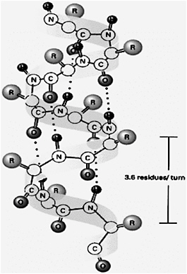
PAGE 192 Right-handed ?-helix.

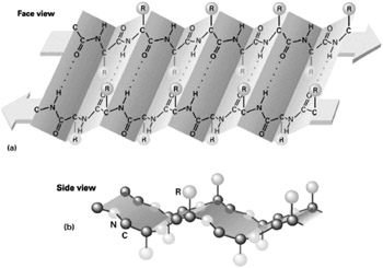
PAGE 193 Diagram sketch of beta-pleated sheat.
(a) View from the top and (b) view from the side.

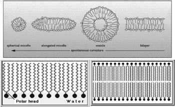
PAGE 215 Various structures that can be formed when hydrophobic phospholipids are free in an aqueous medium.

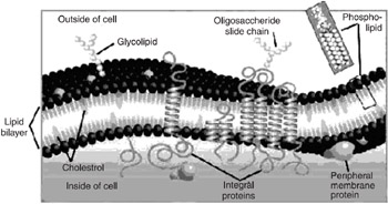
PAGE 215 Fluid mosaic model of a typical Plasma membrane.

PAGE 254 Fluid mosaic model for cell membranes.

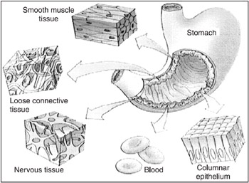
PAGE 264 Various types of tissues present in the stomach.

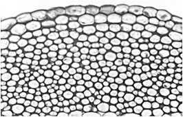
PAGE 269 Collenchyma.

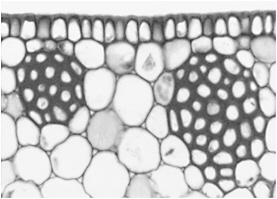
PAGE 269 Sclerenchyma.

PAGE 286 The stages of mitosis.

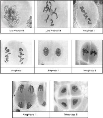
PAGE 287 The Stages of Meiosis (The photographs show meiosis in Lilium longiformum (lilly). Note that as in mitosis the process is dynamic and that the terms do not refer to static stages.

PAGE 288 A diagram representation comparing mitosis and meiosis.

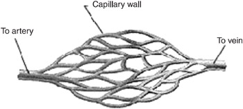
PAGE 304 Capillary beds.

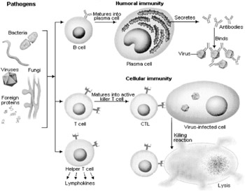
PAGE 322 Pathway of immune response.

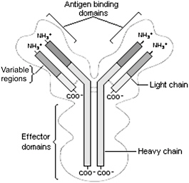
PAGE 323 Structure of an antibody.

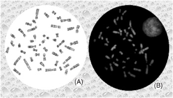
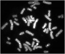
PAGE 470-471 (A) G-banded metaphase cell at 1000x;
(B) Q-banded metaphase cell at 1000x.

PAGE 474 Fish-painted chromosomes.

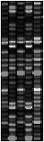
PAGE 609 A sequencing gel after electrophoresis, illuminated with ultra-violet laser. Here, the primers are not radiolabeled. Instead, each type of ddNTPs are fluorescently labeled and therefore are visible in the gel itself under UV. This can be read automatically with a UV laser scanner or reader. The introduction of fluorescent labeling and the automatic reading of the bands became an integral part of the automatic DNA sequencing machine or the automated DNA sequencer, which made the technique of DNA...

UNLIMITED FREE
ACCESS
TO THE WORLD'S BEST IDEAS

SUBMIT
Already a GlobalSpec user? Log in.

This is embarrasing...

An error occurred while processing the form. Please try again in a few minutes.

Customize Your GlobalSpec Experience

Category: DNA Synthesizers
Finish!
Privacy Policy

This is embarrasing...

An error occurred while processing the form. Please try again in a few minutes.