
Products & Services
See also: Categories | Featured Products | Technical Articles | More Information-
Supplier: Flexbar Machine Corporation
Description: shank 1/2" diameter, eyepiece angle 60°. Field of view .100" diameter, focal length 5/8". Optional Edge Block to locate the edge of any work piece to the spindle axis, and V-Block Gage for precision centering of a V-Block to the spindle for cross axis boring. Model No. 17272, Centering
-
Supplier: Titan Tool Supply, Inc.
Description: The Titan Centering Microscopes are designed for job set-up and inspection with vertical milling machines, large jig borers, boring mills, lathes, electrical discharge machines, tape-controlled machines, drill presses. Three Coordinate Measuring Machines. Titan Centering
- Application: Measuring / Toolmaker
-
Supplier: Titan Tool Supply, Inc.
Description: The Titan Centering Microscopes are designed for job set-up and inspection with vertical milling machines, large jig borers, boring mills, lathes, electrical discharge machines, tape-controlled machines, drill presses. Three Coordinate Measuring Machines. Titan Centering
- Total Magnification: 10 to 400 X
- Application: Measuring / Toolmaker
- Features: Monocular
-
-
Supplier: CIQTEK Co., Ltd
Description: CIQTEK Quantum Diamond Microscope (QDM) is a wide-field magnetic resonance based on the principle of spin magnetic resonance in the diamond nitrogen-vacancy center (NV center). The spin quantum state of the NV center luminescence defects is susceptible to the surrounding microwave and static
- Application: Biological / Life Science, Semiconductor Inspection
- Remote Interface: Computer Interface, Image Analysis Processing Software
- Features: Digital Display, Mechanical Stage, Other
- Grade: Research
-
Supplier: CIQTEK Co., Ltd
Description: CIQTEK Scanning NV Microscope Diamond III/IV is a scanning NV magnetometer based on the diamond nitrogen-vacancy center (NV center) and AFM scanning magnetic imaging technology. The sample's magnetic properties are obtained quantitatively and non-destructively by quantum control and readout
- Application: Biological / Life Science, Semiconductor Inspection
- Remote Interface: Computer Interface, Image Analysis Processing Software
- Features: Digital Display, Mechanical Stage, Other
- Grade: Research
-
Supplier: Flexbar Machine Corporation
Description: CENTERING MICROSCOPE Adapts to any machine spindle with maximum accessibility to the operator. 45X magnification for optical position accuracy of .0001". Single dash line reticle for easy observation of the work reference line. Unique compensation of spindle runout by adjustment screw.
-
Supplier: Flexbar Machine Corporation
Description: Standard 1.0X Objective. Model No. 20768, Flexbar K Series Zoom Stereo Microscope Zoom range at 1:5.2 ratio designed on self-centering dovetail ring mechanism, total parfocality and absolute centering
-
Supplier: Newport MKS
Description: Designed as a reverse Casegrain, Newport Reflective Microscope Objectives focus light using reflective surfaces that are broadband coated with a usable wavelength range from 200 nm to 20 µm. Wide spectral band Long working distances High numerical apertures Zero chromatic aberration
- Magnification: 15 to 36 X
- Numerical Aperture: 0.4 to 0.52 NA
- Lens Type: Objective Lens
-
Supplier: Evident Scientific
Description: The AL120 wafer handler series transfers both silicon and compound semiconductor wafers from the cassette to the microscope stage with enhanced capabilities and flexibility, while maintaining an ergonomic design. Accommodates Multiple Wafer Sizes The ability to accommodate different wafer
- Grade: Benchtop
- Eyepiece Style: Binocular
- Microscope Type: Compound
- Features: Digital Display, Mechanical Stage
-
Supplier: Flexbar Machine Corporation
Description: V-BLOCK FOR CENTERING MICROSCOPE Adapts to any machine spindle with maximum accessibility to the operator. 45X magnification for optical position accuracy of .0001". Single dash line reticle for easy observation of the work reference line. Unique compensation of spindle runout by
-
Supplier: Titan Tool Supply, Inc.
Description: A simple pocket microscope with unusual sharpness of definition and excellent lens quality not usually found in small pocket units. The magnification varies from 16X to 32X with a field of view of .118" to .236". The unit has a built-in magnification changer and fine focusing attachment. This
- Total Magnification: 16 to 32 X
- Application: Measuring / Toolmaker
- Features: Monocular
-
Supplier: Evident Scientific
Description: The IX53 has been designed from the ground up to be the finest microscope available for routine inverted microscopic analysis. The combination of excellent optical performance and mechanical quality results in a microscope system of outstanding value and comfort for regular use.
- Working Distance: 27 to 73.3 mm
- Grade: Benchtop
- Eyepiece Style: Binocular
- Application: Biological / Life Science
-
Supplier: Parco Scientific Company
Description: The SBH-6100 series is engineered to meet the state of the art needs of progressive laboratories and classrooms. This microscope features many control functions to allow precise optical clarity. It has rugged construction and high optical standards at an affordable price. Limited lifetime
- Eyepiece Style: Binocular
- Application: Biological / Life Science
- Grade: Student
-
Supplier: Parco Scientific Company
Description: Designed to meet the demands in education and training environments, the new 2000 series with infinity optical system will deliver the best performance for a variety of applications. Limited lifetime warranty. strong>Features: ul style="border-width: 0px; margin: 18px 0px 18px 2em; padding: 0px;
- Eyepiece Style: Binocular
- Application: Biological / Life Science
- Grade: Student
-
Supplier: IntelLiDrives, Inc.
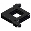
Description: Open-frame XY stages designed with a low profile for a wide range of automated accurate positioning in microscope-based applications. The drive mechanism located on the side of the unit and offers a clear unobstructed two axes aperture to enable light or objects to pass through the center of
-
Supplier: Evident Scientific
Description: Simplify Your Technical Cleanliness The cleanliness of components and parts is at the center of the manufacturing process. Meeting high standards for counting, analyzing, and classifying the often micron-sized contaminant and foreign particles is important for all processes: development,
- Resolution: 100 nm
- Grade: Benchtop
- Remote Interface: Computer Interface, Image Analysis Processing Software
- Features: Digital Display, Fine Focus, Mechanical Stage
-
Supplier: Nikon Metrology
Description: precentered fiber illuminator provides superior epi-fluorescence observation. Lamp centering and focus adjustment are not necessary, even after lamp replacement. The light source can be placed away from the microscope, reducing heat near the microscope and preventing defocusing.
- Total Magnification: 1 to 150 X
- Grade: Benchtop
- Remote Interface: Computer Interface, Image Analysis Processing Software
- Features: Digital Display, Fine Focus, Mechanical Stage
-
Supplier: Parco Scientific Company
Description: Innovative design with durability in mind. Superior optics make it suitable for medical, dental, veterinary use and research settings. This series highlights a smooth operating quintuple reverse nosepiece for easy access to the specimen and stage. Suitable education instrument from High School
- Application: Biological / Life Science
- Grade: Student
- Eyepiece Style: Trinocular
-
Supplier: Edmund Optics Inc.
Description: Simple C-Mount Connection to Infinity Corrected Objectives Compact Design for Simple Integration Into Systems 2/3 Max Sensor Size 14mm In-line Illumination Port M26 x 0.706mm Objective Threading The C-Mount Video Microscope Unit connects an M26 x 0.706mm thread Infinity Corrected Objective to
- Features: Digital Display
- Microscope Type: Fluorescent / UV
-
Supplier: Prior Scientific, Inc.
Description: Key Features: Screen provides positional feedback for all axis Buttons allow for fast up, fast down Z focus Manual focus knob The Interactive Control Center (ICC) is an ergonomically designed multi-functional manual controller for the ProScan™ III microscope automation system. The ICC
- Number of Axes: 3-Axis Control
- Grip Style: Ball
- Mounting: Desktop
- Joystick Application: Precision Joystick Controller
-
Supplier: Newport MKS
Description: The 50105-02 Reflective Microscope Objective is a 15x magnification Reflective Microscope Objective with a back focal length of infinity. Newport Reflective Microscope Objectives are reverse Cassegrains following the Schwarzschild design. Accordingly, they have zero chromatic
- Lens Features: Microoptic
- Features: Other
-
Supplier: Newport MKS
Description: The 50102-02 Reflective Microscope Objective is a 36x magnification Reflective Microscope Objective with a back focal length of infinity. Newport Reflective Microscope Objectives are reverse Cassegrains following the Schwarzschild design. Accordingly, they have zero chromatic
- Lens Features: Microoptic
- Features: Other
-
Supplier: Newport MKS
Description: The 50105-01 Reflective Microscope Objective is a 15x magnification Reflective Microscope Objective with a back focal length of 160 mm. Newport Reflective Microscope Objectives are reverse Cassegrains following the Schwarzschild design. Accordingly, they have zero chromatic
- Focal Length: 160.00000000000003 mm
- Lens Features: Microoptic
- Features: Other
-
Supplier: Titan Tool Supply, Inc.
Description: These compact units have their own light source located in a simple powerful pen light which is adjustable on a two-way swivel. The base is detachable by unscrewing and can be removed for special mountings on production lines, etc. The Titan 70 Series Measuring Microscope has a precision
- Magnification: 10 to 20 X
- Type / Mounting: Arm / Boom / Clamp
- Applications: Assembly, Electronics Manufacturing, Industrial Inspection
- Features: Illuminated
-
Supplier: Qioptiq
Description: The Manual Measuring Stage is primarily used on measuring microscopes and profile projectors in both reflected and transmitted light applications. It can be used with various microscope types from Leica, Nikon, Olympus and Zeiss. Transmitted light measurings due to glass plate on top
- Maximum X-Distance: 0 to 25 mm
- Maximum Y-Distance: 0 to 25 mm
- Minimum Step Size (Resolution): 5 µm
- Features: Graduated / Measuring
-
Supplier: MTI Instruments Inc.
Description: These in-situ miniature bend, fatigue, strain, and tensile testers are designed for use in Scanning Electron Microscopes (SEMs), and Light Microscopes (LMs). The dual leadscrew design symmetrically loads samples while keeping them centered within the scopes field of view. 100, 1000 and
- Maximum X-Distance: 34.00000000000001 mm
- Stage Type: Motorized Stage
-
Supplier: IntelLiDrives, Inc.
Description: Open-frame XY stages designed with a low profile for a wide range of automated accurate positioning in microscope-based applications. The drive mechanism located on the side of the unit and offers a clear unobstruced two axes aperture to enable light or objects to pass through the center of
-
Supplier: IntelLiDrives, Inc.
Description: Open-frame XY stages designed with a low profile for a wide range of automated accurate positioning in microscope-based applications. The drive mechanism located on the side of the unit and offers a clear unobstruced two axes aperture to enable light or objects to pass through the center of
-
Supplier: IntelLiDrives, Inc.
Description: Open-frame XY stages designed with a low profile for a wide range of automated accurate positioning in microscope-based applications. The drive mechanism located on the side of the unit and offers a clear unobstruced two axes aperture to enable light or objects to pass through the center of
-
Supplier: Navitar, Inc.
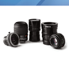
Description: Navitar's Platinum, Raptar and Raptar Pro series of large format lenses meet the demands for high center to edge resolution, low distortion, and application specific F-numbers. These lenses are not modified video lenses; they have been designed to match performance abilities of high-end megapixel
-
Supplier: Edmund Optics Inc.
Description: available to block optical paths, or to create customized mounts. Each blank disk features a 3mm diameter x 1mm deep centering hole for easy customization. Compatible with #85-641 30mm Side Access Plate, #85-717 25mm Interchangeable Optical Mount is equipped with a handle for easy insertion
- Lens Type: Objective Lens










Find Suppliers by Category Top
Featured Products Top
-
 precision in photonics, semiconductor, and microscopy applications. A user programmable virtual pivot point (center of rotation) and high-speed alignment routines built directly into the digital controller firmware further enhance versatility, accelerating setup and throughput. High (read more)
Browse Microscope Stages Datasheets for PI (Physik Instrumente) L.P. -
Microscope XY Stage Democratizes Piezo Motor Advantages: Low Profile, 10nm Resolution, High (read more)
Browse Linear Slides and Linear Stages Datasheets for PI (Physik Instrumente) L.P. -
Leak Test Stations Multiple TIG stations Manufacturing In-Station Flow Hoods XRF Metal Analysis Tool Multiple Glove Boxes and Mechanical Cleaning Tools Hi Power Digital Microscopes Various Machining and (read more)
Browse Electrical Feedthroughs Datasheets for MPF Products, INC. -
astronomical telescopes to diffuse light and help the observer see more distant objects. 2. Microscope: in a microscope, a biconvex lens is used to view thin samples, such as biological sections. 3. Magnifiers: biconvex lenses magnify objects and form an orthogonal (read more)
Browse Optical Lenses Datasheets for Changchun Yutai Optics Co., Ltd. -
Fluorescence Filters for Microscopy and Imaging Fluorescence microscopes and imaging systems utilize fluorescent biomarkers and fluorescence filter sets to create bright, high (read more)
Browse Optical Filters Datasheets for Alluxa, Inc.
Conduct Research Top
-
IFPAC 2006 Under the Microscope
spectroscopic analysis of the core after coating. Two of my favorite talks were by the same person: Fiona Clarke of Pfizer. The first was on using chemical imaging for root cause analysis (RCA). Her talk centered on understanding the process and problems, not just solving why a product failed The concepts were
-
Measuring Weld Throat Thickness with OLYMPUS Stream Image Analysis Software
After metal sheets are welded together, the strength of the welds must be checked to help ensure the integrity of final products. Welds need to have sufficient throat thickness-the distance from the center of the weld face to its root-to meet strength requirements. Whether you are inspecting welds
-
EETimes.com | Electronics Industry News for EEs & Engineering Managers
Philips reportedly plans to sell its stake in ASML Microchip plans three more design centers in China Infineon plans $1 billion move to 300-mm production in Dresden Lara Technology becomes Lara Networks, focusing on Internet infrastructure Foundries tackle rising costs of low-volume runs
-
FDA on Career Applicant Pool: What You Need to Know
for the Holidays Current Trends in GMP Warning Letters Tufts Center for the Study of Drug Development's "Outlook 2005 " Podcast: FDA's Dr. Janet Woodcock on New Directions for FDA in 2007 Pharmaceutical Six Sigma Quality by Design Recession Perception Invades Executive Job Market, Pockets of Growth Emerge
-
Piezo Motors: Principle of Operation for Engineers
and negative charge centers in the material takes place, which then results in an external electrical field. When reversed, an outer electrical field either stretches or compresses the piezoelectric material. The piezoelectric effect is very useful within many applications that involve the production
-
Designing Atmospheric-Pressure Plasma Sources for Surface Engineering of Nanomaterials
microscope glass slides after treating with. air plasma for 2 min. 3.2 Cell preparation. For these experiments, established human cell lines that. represent the broad spectrum of breast cancer disease were. used. As a model for non-transformed cells, MCF10A (ATCC,. Manassas, VA, USA) cells were chosen
-
A Microfluidic System for Biological Particle Enrichment Using Contactless Dielectrophoresis
microscope. slides and the PDMS replica were bonded (Fig. 1F) after exposure. to air plasma for 40 s in a plasma cleaner (PDC-001,. Harrick Plasma, Ithaca, NY). An SEM image of the trapping. zone of the device replica on the silicon master is shown in. Figure 1G. Figure 1H shows the fabricated
More Information Top
 Indicates content that may require registration and/or purchase.

-
Mouse Models for Drug Discovery
Centering microscope with 20X magnification (David Kopf, Model 1915).
-
Sample Preparation Handbook for Transmission Electron Microscopy
Centering Microscope .
-
Quantitative stray field imaging of a magnetic vortex core
Here we use a scanning nitrogen-vacancy (NV) center microscope to quantitatively map the stray magnetic field above a 1 µm-diameter disk of permalloy, unambiguously revealing the vortex core.
-
Alignment of a Spectrometer for Soft X-Ray Laser Diagnostics
A long -working- distance centering microscope is pointed at each pinhole.
-
NV-center imaging of bubble domains in a 6 Å film of cobalt with
perpendicular magnetization
We employ a scanning NV- center microscope to perform stray field imaging of bubble magnetic domains in a perpendicularly magnetized Pt/Co/AlOx trilayer with 6 ˚A of Co.
-
Design of a micro-rotative actuator test bed
Centering microscope .
-
Some Instruments For Quality Assurance In Component Manufacture
The means used to detect the centring error, so that the operator knows how to move the lens on its chuck, vary from simple visual inspection of images formed by reflection at the lens surfaces, to purpose made centring microscopes .
-
Decreasing pore formation in multiple-sheet laser joining with interfacial polymeric contaminations
The extent of pore formation is evaluated by longitudinal (paral- lel to welding direction=y-direction) micro-sections in the weld center ( microscope Axio Imager A1m, Zeiss).
-
Controlled induction of human pancreatic progenitors produces functional beta‐like cells in vitro
Spheres were then trans- ferred to 4°C for approximately 18 h, followed by standard process- ing and analysis by the Electron Microscope Lab/Diabetes Center Microscope Core.