2.9. TWYMAN–GREEN INTERFEROGRAMS AND THEIR ANALYSIS
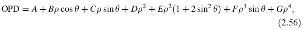
The interferograms due to the primary aberrations can be described by using the
wavefront function by Kingslake (1925–1926), which is given by
where these coefficients represent.
| | A
B
C
D
E
F
G
| Constant (piston) term
Tilt about the y axis
Tilt about the x axis
Reference sphere change, also called defocus
Sagittal astigmatism along the y axis
Sagittal coma along the y axis
Primary spherical aberration |
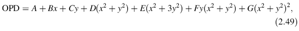
In polar coordinates (ρ, θ), Eq. 2.36 can also be written (x = ρ cos θ; y = ρ sin θ) as
This expression is designed to represent the wavefronts produced in the presence
of the primary aberrations of a centered lens whose point source and image are
displaced in the y direction. Thus, the wavefront is always symmetric about the y axis.
Also, the coma and astigmatism terms are referred to the Petzval surface, which is not
of a great relevance in most interferograms. When testing an optical surface or a
descentered system no symmetry can, in general, be assumed and a more general
wavefront representation has to be considered.
Additionally, it is convenient for the mathematical analysis that the average tilt of
all aberrations is zero with the exception of the two tilts. This is equivalent to
selecting the optimum tilts of the reference wavefront for each aberration. Also,
the average curvature of all aberrations must be zero for all aberrations, with the
exception of the spherical curvature, also called defocus. This is equivalent to
selecting the optimum value of the focus setting for each aberration. These
aberrations are the Zernike polynomials to be described with detail in Chapter 13.
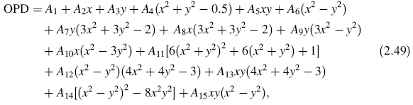
In terms of these aberrations, the wavefront shape up to the fourth order terms can be
written as
or in polar coordinates
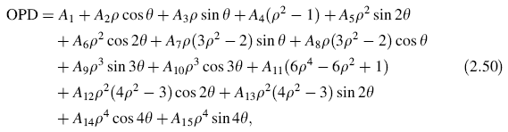
where
| | A1
| Constant (Piston) term
|
| A2 | Tilt about the y axis |
| A3 | Tilt about the x axis |
| A4 | Spherical term, also called defocus |
| A5 | Astigmatism with axis at ± 45o |
| A6 | Astigmatism with axis a at 0o or 90o |
| A7 | Third order coma along y axis |
| A8 | Third order coma along x axis |
| A9 | Triangular astigmatism with base parallel to x axis |
A10
| Triangular astigmatism with base parallel to y axis |
| A11 | Primary spherical aberration |
| A12 | High order astigmatism at 0o or 90o |
| A13 | High order astigmatism at ± 45o |
| A14 | Quadrangular (ashtray) astigmatism 0o or 90o |
| A15 | Quadrangular (ashtray) astigmatism at ± 45o. |
In computing interferograms, a normalized entrance pupil with unit semidiameter
ρ can be assumed. The great advantage of this normalization is that a value of all the
aberration coefficients will represent the same maximum wavefront deformation at
the edge of the pupil.
The relative simplicity of the Kingslake expression allows us an easy and intuitive
analysis of the interferograms, as we will see with some examples. The interferograms
for some aberrations were simulated by calculating the irradiance at a
two-dimensional array of points. A wavelength equal to 632.8 nm was used in these
interferograms, the pupil diameter is 20.0 mm but the values of the coefficients are
defined for a normalized pupil (ρ = 1).
1. Perfect lens. The patterns for a perfect lens without tilts (B = C = 0) and with
tilt (B = 5.0 × 10⁻³) are shown in Figures 2.42(a,b). A perfect lens with defocusing
(D = 3.0 × 10⁻³) and with defocusing and tilt (D = 3.0 × 10⁻³; B = 5.0 × 10⁻³) is
illustrated in Figures 2.42(c,d).
2. Spherical aberration. The patterns for pure spherical aberration were computed
assuming that G = 5.0 × 10⁻³. They are shown at the paraxial focus (D = 0),
without tilts (B = C = 0) and with tilt (B = 5.0 × 10⁻³) in Figures 2.43(a,d). The
patterns at the marginal focus are obtained by setting in Eq. (2.43), only A and D
different from zero,
| |  |

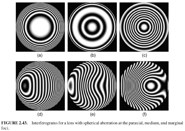
Therefore, we set the defocusing coefficient B = -5.0 × 10⁻³ and the spherical aberration
coefficient G = 5.0 × 10⁻³. These interferograms without (B = C = 0) and with (B = 5.0 × 10⁻³)
tilt are shown in Figures 2.43(c,f). The fringe patterns at the medium focus with B = -10.0 × 10⁻³
are in Figures 2.43(b,e).
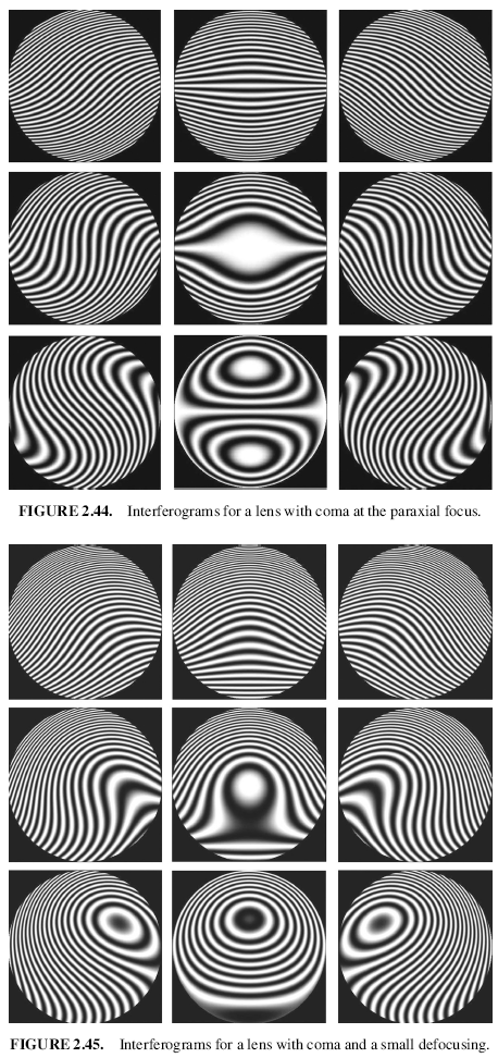
3. Coma. All the patterns for coma were obtained using F = 5.0 × 10⁻³.
Figure 2.44 shows them for the paraxial focus (D = 0) and Figure 2.45 with a small
defocusing (D = 5.0 × 10⁻³). In both figures the central pattern has no tilt
(E = F = 0) and the surrounding pictures are for different tilt combinations
(B = ±5.0 × 10⁻³; C = ±5.0 × 10⁻³).
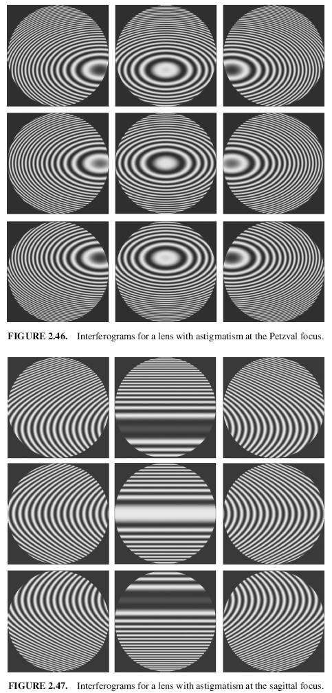
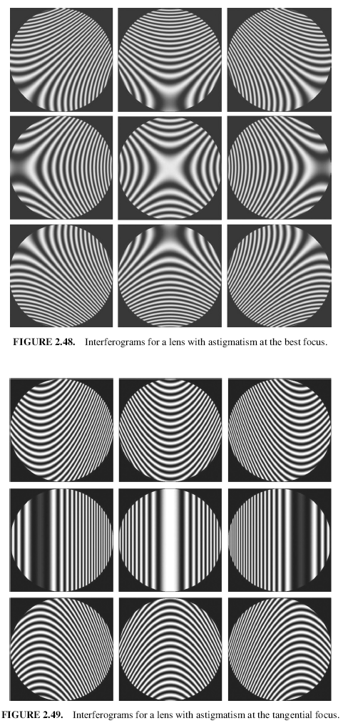
4. Astigmatism. All the patterns for astigmatism were computed for
C = 3.0 × 10⁻³. If = 0, we obtain the Petzval focus. The OPD for astigmatism
can be written from Eq. (2.36) as
| |  |
Therefore, the sagittal focus is obtained for D + E = 0 end the tangential focus for
D + 3E = 0. The medium focus is obtained for D + E = -(D + 3E); hence
D = -2E.
Figure 2.46 shows the patterns at the Petzval focus with tilts in all directions
(B = ±5.0 × 10⁻³; C = ±5.0 × 10⁻³). Figures 2.47–2.49 show the patterns at the
sagittal, medium, and tangential foci, respectively, also with tilts in all directions.
| |  |
| |  |
| |  |
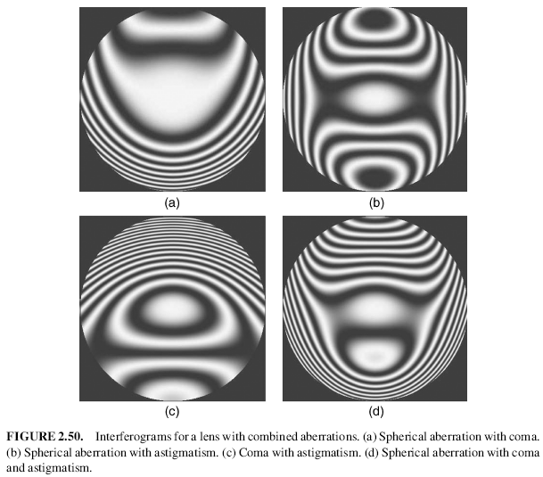
5. Combined Aberrations. Figure 2.50 shows the patterns for combined aberrations:
spherical aberration plus coma (G = 2.0 × 10⁻³ and F = 3.0 × 10⁻³) in
Figure 2.50(a), spherical aberration plus astigmatism (G = 4.0 × 10⁻³ and
E = -2.0 × 10⁻³) in Figure 2.50(b), coma plus astigmatism (F = -2.0 × 10⁻³,
E = 4.0 × 10⁻³) in Figure 2.50(c), and, finally, spherical aberration
plus coma plus astigmatism (G = 5.0 × 10⁻³,F = -2.0 × 10⁻³,E = 4.0 × 10⁻³) in Figure 2.50(d).
Pictures of typical interferograms are shown in a paper by Marechal and Dejonc
(1950). These interferograms can be simulated by beams of fringes of equal inclination
on a Michelson interferometer (Murty, 1960) using the OPDs introduced by a
plane parallel plate and cube corner prisms instead of mirrors or by electronic circuits
on a CRT (Geary et al., 1978 and Geary, 1979).
This type of interferogram was first analyzed by Kingslake (1926–1927). He
measured the OPD at several points on the x and y axes just by fringe counting.
Then, solving a system of linear equations, he computed the OPD coefficients B, C,
D, E, F, G. Another method for analyzing a Twyman–Green interferogram was
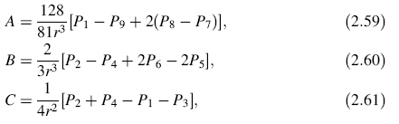
proposed by Saunders (1965). He found that the measurement of four appropriately
chosen points is sufficient to determine any of the three primary aberrations. The
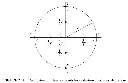
points were selected as in Figure 2.51 and then the aberration coefficients were
computed as
where P1 is the interference order at point i.
If a picture of the interferogram is not taken, the aberration coefficients can be
determined by direct reading on the interferogram setting, looking for interference
patterns with different foci and tilts (Perry, 1923-1924). To make these readings
easier, some optical arrangements may be used to separate symmetrical and asymmetrical
wavefront aberrations as shown by Hariharan and Sen (1961).
© 2007
 Since the publication of the second edition of this book, many important advances
have taken place in the field of optical testing. On one hand, the requirements for
faster and more precise tests are stronger than ever; on the other hand, the new
technological tools permit us to do these tasks much better than before. The need to
describe these advances in this book would lead us to a thicker and hence more
expensive book. This was not compatible with our desire to keep the price as low as
possible, and therefore several new things had to be done. One of them was to reduce
the description of some of the most mathematical sections in the book so as leaving
space for some more applied subjects. Another modification was to reduce as much
as possible the number of references at the end of each chapter, leaving only the most
relevant ones. To compensate it, a CD with the complete and almost exhaustive list of
references is included in the book. Another advantage of this is that the full list of
references is properly classified by topics or its possible applications. Since many
publications may have two, three or more subjects, it is included in each of these
sections. For example, a publication may describe a test that is useful for testing flats,
spheres, and prisms. In that case, this publication is present in all of these sections. A
reader with a particular optical tests need, may find some help by using this reference
list in PDF format. The list of publications in optical testing is so large that it is
impossible to expect that no important reference is missing. If so, the Editor
apologizes for overlooking any important reference. Of course the list may be
updated every one or two years.
Since the publication of the second edition of this book, many important advances
have taken place in the field of optical testing. On one hand, the requirements for
faster and more precise tests are stronger than ever; on the other hand, the new
technological tools permit us to do these tasks much better than before. The need to
describe these advances in this book would lead us to a thicker and hence more
expensive book. This was not compatible with our desire to keep the price as low as
possible, and therefore several new things had to be done. One of them was to reduce
the description of some of the most mathematical sections in the book so as leaving
space for some more applied subjects. Another modification was to reduce as much
as possible the number of references at the end of each chapter, leaving only the most
relevant ones. To compensate it, a CD with the complete and almost exhaustive list of
references is included in the book. Another advantage of this is that the full list of
references is properly classified by topics or its possible applications. Since many
publications may have two, three or more subjects, it is included in each of these
sections. For example, a publication may describe a test that is useful for testing flats,
spheres, and prisms. In that case, this publication is present in all of these sections. A
reader with a particular optical tests need, may find some help by using this reference
list in PDF format. The list of publications in optical testing is so large that it is
impossible to expect that no important reference is missing. If so, the Editor
apologizes for overlooking any important reference. Of course the list may be
updated every one or two years.