Introduction to Applied Statistical Signal Analysis: Guide to Biomedical and Electrical Engineering Applications, Third Edition

The use of envelopes and kernel functions has become more prevalent in the biomedical engineering literature within the last decade the former in the use of image creation and the latter in the creation of continuous time domain signals from point events. Both methods are very different but are placed together in the same chapter because they both create a low-frequency signal from a high-frequency one.
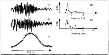
For some biomedical phenomena, not only is the direct measurement important but also its envelope. Two examples will illustrate the importance. Consider the investigation of uterine physiology shown in Figure 10.1 (Duchene, 1995). Figures 10.1a and b show the muscular activity in the form of the electromyogram (EMG) and Figure 10.1c shows the intrauterine pressure (IUP). Often it is desired to estimate the latter from the envelope of the EMG signal. This is because the EMG is a measurement of the electrical activity that occurs during the excitation-contraction coupling of muscle. As one can see, the IUP is low in frequency content and is approximated by the envelope of the EMG, and its peak is delayed in time with respect to the peak of the EMG. In addition, the instantaneous frequency of the EMG may be important as well (see Figure 10.2b). This is an estimate of the major frequency component as a function of time.